File:Design and characterization of a wearable cardiac imager.webp

Page contents not supported in other languages.
This is a file from the Wikimedia Commons
From Wikipedia, the free encyclopedia

Original file(2,157 × 2,120 pixels, file size: 428 KB, MIME type: image/webp)

Summary

Description
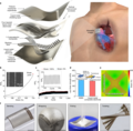
English: "a, Schematics showing the exploded view of the wearable imager, with key components labelled (left) and its working principle (right). b, Resistance of the liquid metal composite electrode as a function of uniaxial tensile strain. The electrode can be stretched up to about 750% without failure. The y axis is the relative resistance defined as R/R0, in which R0 and R are the measured resistances at 0% strain and a given strain, respectively. The inset is a scanning electron micrograph of the liquid metal composite electrodes with a width as small as about 30 µm. Scale bar, 50 μm. c, Cycling performance of the electrode between 0% and 100% uniaxial tensile strain, showing the robustness of the electrode. The inset shows the zoomed-in features of the graph during cyclic stretching and relaxing of the electrode. d, Lap shear strength of the bonding between transducer elements and SEBS or liquid metal composite electrode. Data are mean and s.d. from n = 3 tests. The inset is a schematic setup of the lap shear test. e, Finite element analysis of the entire device under 110% biaxial stretching. f, Optical images showing the mechanical compliance of the wearable imager when bent on a developable surface, wrapped around a non-developable surface, poked and twisted. Scale bars, 5 mm."
Date
Source https://www.nature.com/articles/s41586-022-05498-z
Author Authors of the study: Hongjie Hu, Hao Huang, Mohan Li, Xiaoxiang Gao, Lu Yin, Ruixiang Qi, Ray S. Wu, Xiangjun Chen, Yuxiang Ma, Keren Shi, Chenghai Li, Timothy M. Maus, Brady Huang, Chengchangfeng Lu, Muyang Lin, Sai Zhou, Zhiyuan Lou, Yue Gu, Yimu Chen, Yusheng Lei, Xinyu Wang, Ruotao Wang, Wentong Yue, Xinyi Yang, Yizhou Bian, Jing Mu, Geonho Park, Shu Xiang, Shengqiang Cai, Paul W. Corey, Joseph Wang & Sheng Xu

Licensing

w:en:Creative Commons
attribution
This file is licensed under the Creative Commons Attribution 4.0 International license.
You are free:
  • to share – to copy, distribute and transmit the work
  • to remix – to adapt the work
Under the following conditions:
  • attribution – You must give appropriate credit, provide a link to the license, and indicate if changes were made. You may do so in any reasonable manner, but not in any way that suggests the licensor endorses you or your use.

Captions

From the study "A wearable cardiac ultrasound imager"

Items portrayed in this file

depicts

25 January 2023

image/webp

File history

Click on a date/time to view the file as it appeared at that time.

Date/TimeThumbnailDimensionsUserComment
current21:39, 22 February 2023Thumbnail for version as of 21:39, 22 February 20232,157 × 2,120 (428 KB)PrototyperspectiveUploaded a work by Authors of the study: Hongjie Hu, Hao Huang, Mohan Li, Xiaoxiang Gao, Lu Yin, Ruixiang Qi, Ray S. Wu, Xiangjun Chen, Yuxiang Ma, Keren Shi, Chenghai Li, Timothy M. Maus, Brady Huang, Chengchangfeng Lu, Muyang Lin, Sai Zhou, Zhiyuan Lou, Yue Gu, Yimu Chen, Yusheng Lei, Xinyu Wang, Ruotao Wang, Wentong Yue, Xinyi Yang, Yizhou Bian, Jing Mu, Geonho Park, Shu Xiang, Shengqiang Cai, Paul W. Corey, Joseph Wang & Sheng Xu from https://www.nature.com/articles/s41586-022-05498-z wit...
The following pages on the English Wikipedia use this file (pages on other projects are not listed):