
File:Echinococcus vogeli.jpg
Size of this preview: 800 × 526 pixels. Other resolutions: 320 × 210 pixels | 640 × 421 pixels | 1,024 × 673 pixels | 1,280 × 841 pixels | 3,059 × 2,010 pixels.
Original file (3,059 × 2,010 pixels, file size: 1.34 MB, MIME type: image/jpeg)
| This is a file from the Wikimedia Commons. Information from its description page there is shown below. Commons is a freely licensed media file repository. You can help. |
Summary
| Description |
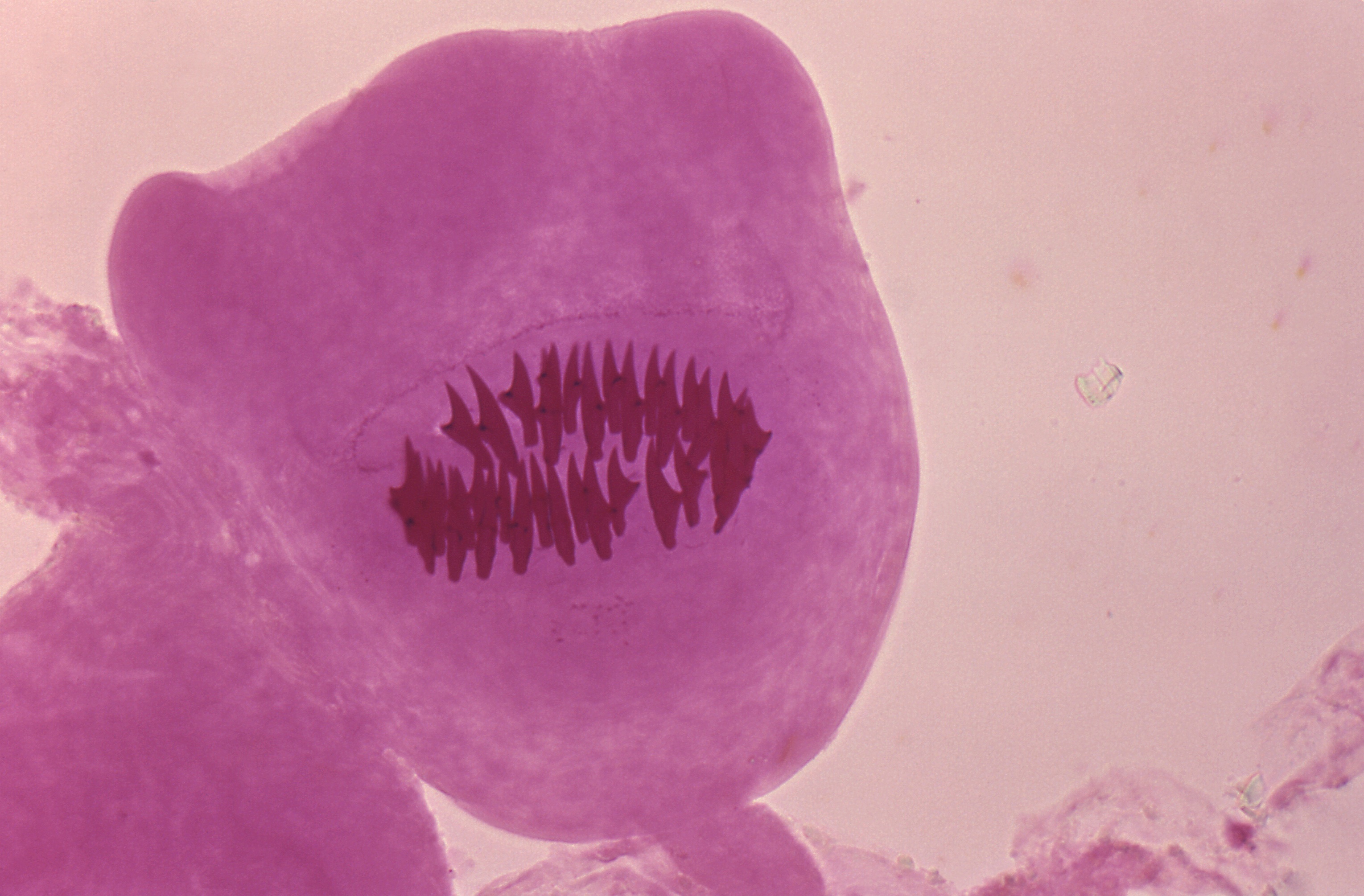
English: This photomicrograph of a tissue sample harvested from a cyst, which was harbored inside a gorilla, revealed the presence of these protoscolex hooks of a microscopic Echinococcus vogeli tapeworm. The larval stage of this microscopic tapeworm is one of the causative agents of alveolar hydatid disease (AHD), an infection in humans that causes parasitic tumors to form, mainly in the liver, but can also appear in other organs as well. |
||
| Date | |||
| Source |
|
||
| Author |
|
||
| Permission (Reusing this file) |
PD-USGov-HHS-CDC English: None - This image is in the public domain and thus free of any copyright restrictions. As a matter of courtesy we request that the content provider be credited and notified in any public or private usage of this image. |
Licensing
This image is a work of the Centers for Disease Control and Prevention, part of the United States Department of Health and Human Services, taken or made as part of an employee's official duties. As a work of the U.S. federal government, the image is in the public domain.
|
Original upload log
| date/time | username | resolution | size | edit summary |
|---|---|---|---|---|
| 11:42, 18 November 2007 | User:Filip em | 700×478 | 30 KB | {{Information |Description=This is a photomicrograph of a Echinococcus vogeli protoscolex taken from a cyst within a gorilla. The larval stage of the microscopic tapeworm Echinococcus vogeli is one of the causative agents of Alveolar Hydatid Disease (AHD |
File history
Click on a date/time to view the file as it appeared at that time.
| Date/Time | Thumbnail | Dimensions | User | Comment | |
|---|---|---|---|---|---|
| current | 07:17, 4 August 2020 | | 3,059 × 2,010 (1.34 MB) | TommyG | Larger version from source |
| 04:30, 17 July 2008 |  | 700 × 478 (30 KB) | BetacommandBot | move approved by: User:Luigi Chiesa This image was moved from Image:PHIL 2893 lores.jpg == Summary == {{Information |Description={{en|This is a photomicrograph of a Echinococcus vogeli protoscolex taken from a cyst within a gorilla. The larval |
File usage
The following pages on the English Wikipedia use this file (pages on other projects are not listed):
Global file usage
The following other wikis use this file:
- Usage on bg.wikipedia.org
- Usage on cs.wikipedia.org
- Usage on de.wikipedia.org
- Usage on fr.wikipedia.org
- Usage on pl.wikipedia.org
- Usage on www.wikidata.org