File:Enterobius vermicularis LifeCycle.gif

Page contents not supported in other languages.
This is a file from the Wikimedia Commons
From Wikipedia, the free encyclopedia

Enterobius_vermicularis_LifeCycle.gif(435 × 497 pixels, file size: 17 KB, MIME type: image/gif)

Description

Enterobiasis [Enterobius vermicularis]

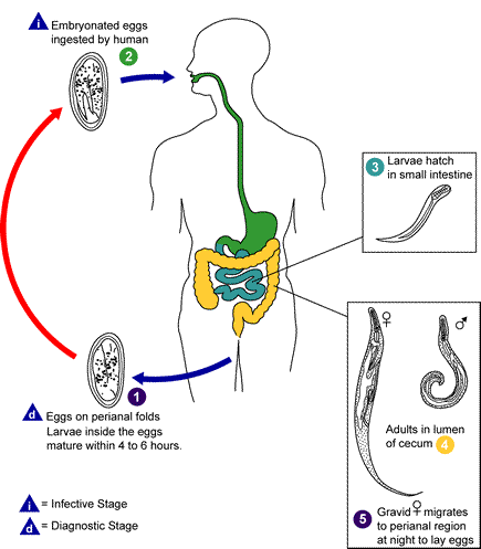
Life cycle of Enterobius vermicularis

1: Eggs are deposited on perianal folds.
2: Self-infection occurs by transferring infective eggs to the mouth with hands that have scratched the perianal area.
3: Person-to-person transmission can also occur through handling of contaminated clothes or bed linens. Enterobiasis may also be acquired through surfaces in the environment that are contaminated with pinworm eggs (e.g., curtains, carpeting). Some small number of eggs may become airborne and inhaled. These would be swallowed and follow the same development as ingested eggs. Following ingestion of infective eggs, the larvae hatch in the small intestine...
4: and the adults establish themselves in the colon.
5: The time interval from ingestion of infective eggs to oviposition by the adult females is about one month. The life span of the adults is about two months. Gravid females migrate nocturnally outside the anus and oviposit while crawling on the skin of the perianal area. The larvae contained inside the eggs develop (the eggs become infective) in 4 to 6 hours under optimal conditions.

1: Retroinfection, or the migration of newly hatched larvae from the anal skin back into the rectum, may occur but the frequency with which this happens is unknown.
Source http://www.dpd.cdc.gov/dpdx/images/ParasiteImages/A-F/Enterobiasis/Enterobius_LifeCycle.gif
Author Centers for Disease Control & Prevention (CDC, http://www.dpd.cdc.gov/dpdx)
Permission
(Reusing this file)
Public domain
This image is a work of the Centers for Disease Control and Prevention, part of the United States Department of Health and Human Services, taken or made as part of an employee's official duties. As a work of the U.S. federal government, the image is in the public domain.

eesti  Deutsch  čeština  español  português  English  français  Nederlands  polski  slovenščina  suomi  македонски  українська  日本語  中文(简体)‎  中文(繁體)‎  العربية  +/−

Other versions

Derivative works of this file:  Enterobius vermicularis LifeCycle-gl.gif

Image:Enterobius vermicularis LifeCycle B.svg (only legends); Image:Enterobius vermicularis LifeCycle pt-version.svg (portuguese version).

File history

Click on a date/time to view the file as it appeared at that time.

Date/TimeThumbnailDimensionsUserComment
current17:27, 15 December 2012Thumbnail for version as of 17:27, 15 December 2012435 × 497 (17 KB)Cartoffelwatermark removed
18:50, 13 May 2006Thumbnail for version as of 18:50, 13 May 2006435 × 497 (21 KB)Patho{{Information| |Description=Enterobiasis [Enterobius vermicularis] Life cycle of Enterobius vermicularis Eggs are deposited on perianal folds. Self-infection occurs by transferring infective eggs to the mouth with hands that have scratched the peria
The following pages on the English Wikipedia use this file (pages on other projects are not listed):

Global file usage

The following other wikis use this file: