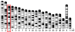
Peptidoglycan recognition protein 4

Peptidoglycan recognition protein 4 (PGLYRP4, formerly PGRP-Iβ) is an antibacterial and anti-inflammatory innate immunity protein that in humans is encoded by the PGLYRP4 gene.[5][6][7][8]
Discovery[edit]
PGLYRP4 (formerly PGRP-Iβ), a member of a family of human Peptidoglycan Recognition Proteins (PGRPs), was discovered in 2001 by Roman Dziarski and coworkers who cloned and identified the genes for three human PGRPs, PGRP-L, PGRP-Iα, and PGRP-Iβ (named for long and intermediate size transcripts),[5] and established that human genome codes for a family of 4 PGRPs: PGRP-S (short PGRP or PGRP-S[9]) and PGRP-L, PGRP-Iα, and PGRP-Iβ.[5] Subsequently, the Human Genome Organization Gene Nomenclature Committee changed the gene symbols of PGRP-S, PGRP-L, PGRP-Iα, and PGRP-Iβ to PGLYRP1 (peptidoglycan recognition protein 1), PGLYRP2 (peptidoglycan recognition protein 2), PGLYRP3 (peptidoglycan recognition protein 3), and PGLYRP4 (peptidoglycan recognition protein 4), respectively, and this nomenclature is currently also used for other mammalian PGRPs.
Tissue distribution and secretion[edit]
PGLYRP4 has similar expression to PGLYRP3 (peptidoglycan recognition protein 3) but not identical.[5][6] PGLYRP4 is constitutively expressed in the skin, in the eye, in the mucous membranes in the tongue, throat, and esophagus, in the salivary glands and mucus-secreting cells in the throat, and at a much lower level in the remaining parts of the intestinal tract.[5][6][10][11] Bacteria and their products increase the expression of PGLYRP4 in keratinocytes and oral epithelial cells.[6][12] Mouse PGLYRP4 is also differentially expressed in the developing brain and this expression is influenced by the intestinal microbiome.[13] PGLYRP4 is secreted and forms disulfide-linked dimers.[6]
Structure[edit]
PGLYRP4, similar to PGLYRP3, has two peptidoglycan-binding type 2 amidase domains (also known as PGRP domains), which are not identical (have 34% amino acid identity in humans)[5][14] and do not have amidase enzymatic activity.[15] PGLYRP4 is secreted, it is glycosylated, and its glycosylation is required for its bactericidal activity.[6] PGLYRP4 forms disulfide-linked homodimers, but when expressed in the same cells with PGLYRP3, it forms PGLYRP3:PGLYRP4 disulfide-linked heterodimers.[6]
The C-terminal peptidoglycan-binding domain of human PGLYRP4 has been crystallized and its structure solved (in a free form and in a complex with peptidoglycan fragment, disaccharide-pentapeptide)[16] and is similar to human PGLYRP1[17] and PGLYRP3.[18][19] PGLYRP4 C-terminal PGRP domain contains central β-sheet composed of six β-strands surrounded by three α-helices and three short helices and N-terminal segment unique to PGRPs and not found in bacteriophage and prokaryotic amidases.[16] PGLYRP4 C-terminal PGRP domain contains three disulfide bonds, one broadly conserved in invertebrate and vertebrate PRGPs, one conserved in all mammalian PGRPs, and one unique to mammalian PGLYRP1, PGLYRP3, and PGLYRP4, but not found in the amidase-active PGLYRP2.[5][15][16][17][18] The structures of the entire PGLYRP4 molecule (with two PGRP domains) and of the disulfide-linked dimer are unknown.
Functions[edit]
The PGLYRP4 protein plays an important role in the innate immune responses.
Peptidoglycan binding[edit]
PGLYRP4 binds peptidoglycan, a polymer of β(1-4)-linked N-acetylglucosamine (GlcNAc) and N-acetylmuramic acid (MurNAc) cross-linked by short peptides, the main component of bacterial cell wall.[5][6][16] PGLYRP4 (its C-terminal PGRP domain) binds peptidoglycan fragment, MurNAc-pentapeptide (MurNAc-L-Ala-γ-D-Gln-L-Lys-D-Ala-D-Ala), with Kd = 1.2 x 10⁻⁵, but similar to PGLYRP3 (and unlike PGLYRP1) does not bind meso-diaminopimelic acid (m-DAP)-containing fragment (MurNAc-L-Ala-γ-D-Gln-DAP-D-Ala-D-Ala).[16] m-DAP is present in the third position of peptidoglycan peptide in Gram-negative bacteria and Gram-positive bacilli, whereas L-lysine is in this position in peptidoglycan peptide in Gram-positive cocci. Thus, PGLYRP4 C-terminal PGRP domain has a preference for binding peptidoglycan fragments from Gram-positive cocci. The fine specificity of the PGLYRP4 N-terminal PGRP domain is not known.
Bactericidal activity[edit]
Human PGLYRP4 is directly bactericidal for both Gram-positive (Bacillus subtilis, Bacillus licheniformis, Bacillus cereus, Lactobacillus acidophilus, Listeria monocytogenes, Staphylococcus aureus) and Gram-negative (Escherichia coli, Proteus vulgaris, Salmonella enterica) bacteria[6][20][21][22][23][24] and is also active against Chlamydia trachomatis.[25]
In Gram-positive bacteria, human PGLYRP4 binds to the separation sites of the newly formed daughter cells, created by bacterial peptidoglycan-lytic endopeptidases, LytE and LytF in B. subtilis, which separate the daughter cells after cell division.[21] These cell-separating endopeptidases likely expose PGLYRP4-binding muramyl peptides, as shown by co-localization of PGLYRP4 and LytE and LytF at the cell-separation sites, and no binding of PGLYRP4 to other regions of the cell wall with highly cross-linked peptidoglycan.[21] This localization is necessary for the bacterial killing, because mutants that lack LytE and LytF endopeptidases and do not separate after cell division, do not bind PGLYRP4, and are also not readily killed by PGLYRP4.[21]
The mechanism of bacterial killing by PGLYRP4 is based on induction of lethal envelope stress, which eventually leads to the shutdown of transcription and translation.[21] PGLYRP4-induced killing involves simultaneous induction of three stress responses in both Gram-positive and Gram-negative bacteria: oxidative stress due to production of reactive oxygen species (hydrogen peroxide and hydroxyl radicals), thiol stress due to depletion (oxidation) of cellular thiols, and metal stress due to an increase in intracellular free (labile) metal ions.[21][22] PGLYRP4-induced oxidative and thiol stress involve malfunction of the respiratory electron transport chain in bacteria.[23][24][26] PGLYRP4-induced bacterial killing does not involve cell membrane permeabilization, which is typical for defensins and other antimicrobial peptides, cell wall hydrolysis, or osmotic shock.[6][20][21] Human PGLYRP4 has synergistic bactericidal activity with antibacterial peptides.[20]
Defense against infections[edit]
PGLYRP4 plays a limited role in host defense against infections. Intranasal administration of PGLYRP4 protects mice from lung infection with S. aureus and E. coli[6][27] and PGLYRP4-deficient mice are more sensitive to Streptococcus pneumoniae-induced pneumonia.[28]
Maintaining microbiome[edit]
Mouse PGLYRP4 plays a role in maintaining healthy microbiome, as PGLYRP4-deficient mice have significant changes in the composition of their intestinal microbiome,[11][28][29] which affects their increased sensitivity to lung inflammation and severity of S. pneumoniae-induced pneumonia.[28]
Effects on inflammation[edit]
Mouse PGLYRP4 plays a role in maintaining anti- and pro-inflammatory homeostasis in the intestine, skin, and lungs. PGLYRP4-deficient mice are more sensitive than wild type mice to dextran sodium sulfate (DSS)-induced colitis, which indicates that PGLYRP4 protects mice from DSS-induced colitis.[11]
PGLYRP4-deficient mice are also more sensitive than wild type mice to experimentally induced atopic dermatitis.[30] These results indicate that mouse PGLYRP4 is anti-inflammatory and protects skin from inflammation. This anti-inflammatory effect in the skin is due to decreased numbers and activity of T helper 17 (Th17) cells and increased numbers of T regulatory (Treg) cells.[30] PGLYRP4-deficient mice also have increased inflammatory responses in the lungs during S. pneumoniae-induced pneumonia associated with impaired bacterial clearance[28] and more severe pulmonary inflammation following Bordetella pertussis infection,[31] indicating anti-inflammatory role of PGLYRP4 in the lungs.
Medical relevance[edit]
Genetic PGLYRP4 variants are associated with some diseases. Patients with inflammatory bowel disease (IBD), which includes Crohn's disease and ulcerative colitis, have significantly more frequent missense variants in PGLYRP4 gene (and also in the other three PGLYRP genes) than healthy controls.[14] PGLYRP4 variants are also associated with Parkinson's disease,[32][33] psoriasis,[34][35] and ovarian cancer.[36] These results suggest that PGLYRP4 protects humans from these diseases, and that mutations in PGLYRP4 gene are among the genetic factors predisposing to these diseases. PGLYRP4 variants are also associated with the composition of airway microbiome.[37]
See also[edit]
- Peptidoglycan recognition protein
- Peptidoglycan recognition protein 1
- Peptidoglycan recognition protein 2
- Peptidoglycan recognition protein 3
- Peptidoglycan
- Innate immune system
- Bacterial cell walls
References[edit]
- ^ a b c GRCh38: Ensembl release 89: ENSG00000163218 – Ensembl, May 2017
- ^ a b c GRCm38: Ensembl release 89: ENSMUSG00000042250 – Ensembl, May 2017
- ^ "Human PubMed Reference:". National Center for Biotechnology Information, U.S. National Library of Medicine.
- ^ "Mouse PubMed Reference:". National Center for Biotechnology Information, U.S. National Library of Medicine.
- ^ a b c d e f g h Liu C, Xu Z, Gupta D, Dziarski R (September 2001). "Peptidoglycan recognition proteins: a novel family of four human innate immunity pattern recognition molecules". The Journal of Biological Chemistry. 276 (37): 34686–34694. doi:10.1074/jbc.M105566200. PMID 11461926. S2CID 44619852.
- ^ a b c d e f g h i j k Lu X, Wang M, Qi J, Wang H, Li X, Gupta D, Dziarski R (March 2006). "Peptidoglycan recognition proteins are a new class of human bactericidal proteins". The Journal of Biological Chemistry. 281 (9): 5895–5907. doi:10.1074/jbc.M511631200. PMID 16354652. S2CID 21943426.
- ^ "PGLYRP4 peptidoglycan recognition protein 4 [Homo sapiens (human)] - Gene - NCBI". www.ncbi.nlm.nih.gov. Retrieved 2020-11-04.
- ^ "PGLYRP4 - Peptidoglycan recognition protein 4 precursor - Homo sapiens (Human) - PGLYRP4 gene & protein". www.uniprot.org. Retrieved 2020-11-04.
- ^ Kang D, Liu G, Lundström A, Gelius E, Steiner H (August 1998). "A peptidoglycan recognition protein in innate immunity conserved from insects to humans". Proceedings of the National Academy of Sciences of the United States of America. 95 (17): 10078–10082. Bibcode:1998PNAS...9510078K. doi:10.1073/pnas.95.17.10078. PMC 21464. PMID 9707603.
- ^ Mathur P, Murray B, Crowell T, Gardner H, Allaire N, Hsu YM, et al. (June 2004). "Murine peptidoglycan recognition proteins PglyrpIalpha and PglyrpIbeta are encoded in the epidermal differentiation complex and are expressed in epidermal and hematopoietic tissues". Genomics. 83 (6): 1151–1163. doi:10.1016/j.ygeno.2004.01.003. PMID 15177568.
- ^ a b c Saha S, Jing X, Park SY, Wang S, Li X, Gupta D, Dziarski R (August 2010). "Peptidoglycan recognition proteins protect mice from experimental colitis by promoting normal gut flora and preventing induction of interferon-gamma". Cell Host & Microbe. 8 (2): 147–162. doi:10.1016/j.chom.2010.07.005. PMC 2998413. PMID 20709292.
- ^ Uehara A, Sugawara Y, Kurata S, Fujimoto Y, Fukase K, Kusumoto S, et al. (May 2005). "Chemically synthesized pathogen-associated molecular patterns increase the expression of peptidoglycan recognition proteins via toll-like receptors, NOD1 and NOD2 in human oral epithelial cells". Cellular Microbiology. 7 (5): 675–686. doi:10.1111/j.1462-5822.2004.00500.x. PMID 15839897. S2CID 20544993.
- ^ Arentsen T, Qian Y, Gkotzis S, Femenia T, Wang T, Udekwu K, et al. (February 2017). "The bacterial peptidoglycan-sensing molecule Pglyrp2 modulates brain development and behavior". Molecular Psychiatry. 22 (2): 257–266. doi:10.1038/mp.2016.182. PMC 5285465. PMID 27843150.
- ^ a b Zulfiqar F, Hozo I, Rangarajan S, Mariuzza RA, Dziarski R, Gupta D (2013). "Genetic Association of Peptidoglycan Recognition Protein Variants with Inflammatory Bowel Disease". PLOS ONE. 8 (6): e67393. Bibcode:2013PLoSO...867393Z. doi:10.1371/journal.pone.0067393. PMC 3686734. PMID 23840689.
- ^ a b Wang ZM, Li X, Cocklin RR, Wang M, Wang M, Fukase K, et al. (December 2003). "Human peptidoglycan recognition protein-L is an N-acetylmuramoyl-L-alanine amidase". The Journal of Biological Chemistry. 278 (49): 49044–49052. doi:10.1074/jbc.M307758200. PMID 14506276. S2CID 35373818.
- ^ a b c d e Cho S, Wang Q, Swaminathan CP, Hesek D, Lee M, Boons GJ, et al. (May 2007). "Structural insights into the bactericidal mechanism of human peptidoglycan recognition proteins". Proceedings of the National Academy of Sciences of the United States of America. 104 (21): 8761–8766. Bibcode:2007PNAS..104.8761C. doi:10.1073/pnas.0701453104. PMC 1885576. PMID 17502600.
- ^ a b Guan R, Wang Q, Sundberg EJ, Mariuzza RA (April 2005). "Crystal structure of human peptidoglycan recognition protein S (PGRP-S) at 1.70 A resolution". Journal of Molecular Biology. 347 (4): 683–691. doi:10.1016/j.jmb.2005.01.070. PMID 15769462.
- ^ a b Guan R, Malchiodi EL, Wang Q, Schuck P, Mariuzza RA (July 2004). "Crystal structure of the C-terminal peptidoglycan-binding domain of human peptidoglycan recognition protein Ialpha". The Journal of Biological Chemistry. 279 (30): 31873–31882. doi:10.1074/jbc.M404920200. PMID 15140887. S2CID 29969809.
- ^ Guan R, Roychowdhury A, Ember B, Kumar S, Boons GJ, Mariuzza RA (December 2004). "Structural basis for peptidoglycan binding by peptidoglycan recognition proteins". Proceedings of the National Academy of Sciences of the United States of America. 101 (49): 17168–17173. Bibcode:2004PNAS..10117168G. doi:10.1073/pnas.0407856101. PMC 535381. PMID 15572450.
- ^ a b c Wang M, Liu LH, Wang S, Li X, Lu X, Gupta D, Dziarski R (March 2007). "Human peptidoglycan recognition proteins require zinc to kill both gram-positive and gram-negative bacteria and are synergistic with antibacterial peptides". Journal of Immunology. 178 (5): 3116–3125. doi:10.4049/jimmunol.178.5.3116. PMID 17312159. S2CID 22160694.
- ^ a b c d e f g Kashyap DR, Wang M, Liu LH, Boons GJ, Gupta D, Dziarski R (June 2011). "Peptidoglycan recognition proteins kill bacteria by activating protein-sensing two-component systems". Nature Medicine. 17 (6): 676–683. doi:10.1038/nm.2357. PMC 3176504. PMID 21602801.
- ^ a b Kashyap DR, Rompca A, Gaballa A, Helmann JD, Chan J, Chang CJ, et al. (July 2014). "Peptidoglycan recognition proteins kill bacteria by inducing oxidative, thiol, and metal stress". PLOS Pathogens. 10 (7): e1004280. doi:10.1371/journal.ppat.1004280. PMC 4102600. PMID 25032698.
- ^ a b Kashyap DR, Kuzma M, Kowalczyk DA, Gupta D, Dziarski R (September 2017). "Bactericidal peptidoglycan recognition protein induces oxidative stress in Escherichia coli through a block in respiratory chain and increase in central carbon catabolism". Molecular Microbiology. 105 (5): 755–776. doi:10.1111/mmi.13733. PMC 5570643. PMID 28621879.
- ^ a b Kashyap DR, Kowalczyk DA, Shan Y, Yang CK, Gupta D, Dziarski R (February 2020). "Formate dehydrogenase, ubiquinone, and cytochrome bd-I are required for peptidoglycan recognition protein-induced oxidative stress and killing in Escherichia coli". Scientific Reports. 10 (1): 1993. Bibcode:2020NatSR..10.1993K. doi:10.1038/s41598-020-58302-1. PMC 7005000. PMID 32029761.
- ^ Bobrovsky P, Manuvera V, Polina N, Podgorny O, Prusakov K, Govorun V, Lazarev V (July 2016). "Recombinant Human Peptidoglycan Recognition Proteins Reveal Antichlamydial Activity". Infection and Immunity. 84 (7): 2124–2130. doi:10.1128/IAI.01495-15. PMC 4936355. PMID 27160295.
- ^ Yang CK, Kashyap DR, Kowalczyk DA, Rudner DZ, Wang X, Gupta D, Dziarski R (January 2021). "Respiratory chain components are required for peptidoglycan recognition protein-induced thiol depletion and killing in Bacillus subtilis and Escherichia coli". Scientific Reports. 11 (1): 64. doi:10.1038/s41598-020-79811-z. PMC 7794252. PMID 33420211.
- ^ Dziarski R, Kashyap DR, Gupta D (June 2012). "Mammalian peptidoglycan recognition proteins kill bacteria by activating two-component systems and modulate microbiome and inflammation". Microbial Drug Resistance. 18 (3): 280–285. doi:10.1089/mdr.2012.0002. PMC 3412580. PMID 22432705.
- ^ a b c d Dabrowski AN, Shrivastav A, Conrad C, Komma K, Weigel M, Dietert K, et al. (2019). "Peptidoglycan Recognition Protein 4 Limits Bacterial Clearance and Inflammation in Lungs by Control of the Gut Microbiota". Frontiers in Immunology. 10: 2106. doi:10.3389/fimmu.2019.02106. PMC 6763742. PMID 31616404.
- ^ Dziarski R, Park SY, Kashyap DR, Dowd SE, Gupta D (2016). "Pglyrp-Regulated Gut Microflora Prevotella falsenii, Parabacteroides distasonis and Bacteroides eggerthii Enhance and Alistipes finegoldii Attenuates Colitis in Mice". PLOS ONE. 11 (1): e0146162. Bibcode:2016PLoSO..1146162D. doi:10.1371/journal.pone.0146162. PMC 4699708. PMID 26727498.
- ^ a b Park SY, Gupta D, Kim CH, Dziarski R (2011). "Differential effects of peptidoglycan recognition proteins on experimental atopic and contact dermatitis mediated by Treg and Th17 cells". PLOS ONE. 6 (9): e24961. Bibcode:2011PLoSO...624961P. doi:10.1371/journal.pone.0024961. PMC 3174980. PMID 21949809.
- ^ Skerry C, Goldman WE, Carbonetti NH (February 2019). "Peptidoglycan Recognition Protein 4 Suppresses Early Inflammatory Responses to Bordetella pertussis and Contributes to Sphingosine-1-Phosphate Receptor Agonist-Mediated Disease Attenuation". Infection and Immunity. 87 (2). doi:10.1128/IAI.00601-18. PMC 6346131. PMID 30510103.
- ^ Goldman SM, Kamel F, Ross GW, Jewell SA, Marras C, Hoppin JA, et al. (August 2014). "Peptidoglycan recognition protein genes and risk of Parkinson's disease". Movement Disorders. 29 (9): 1171–1180. doi:10.1002/mds.25895. PMC 4777298. PMID 24838182.
- ^ Gorecki AM, Bakeberg MC, Theunissen F, Kenna JE, Hoes ME, Pfaff AL, et al. (2020-11-17). "Single Nucleotide Polymorphisms Associated With Gut Homeostasis Influence Risk and Age-at-Onset of Parkinson's Disease". Frontiers in Aging Neuroscience. 12: 603849. doi:10.3389/fnagi.2020.603849. PMC 7718032. PMID 33328979.
- ^ Sun C, Mathur P, Dupuis J, Tizard R, Ticho B, Crowell T, et al. (March 2006). "Peptidoglycan recognition proteins Pglyrp3 and Pglyrp4 are encoded from the epidermal differentiation complex and are candidate genes for the Psors4 locus on chromosome 1q21". Human Genetics. 119 (1–2): 113–125. doi:10.1007/s00439-005-0115-8. PMID 16362825. S2CID 31486449.
- ^ Kainu K, Kivinen K, Zucchelli M, Suomela S, Kere J, Inerot A, et al. (February 2009). "Association of psoriasis to PGLYRP and SPRR genes at PSORS4 locus on 1q shows heterogeneity between Finnish, Swedish and Irish families". Experimental Dermatology. 18 (2): 109–115. doi:10.1111/j.1600-0625.2008.00769.x. PMID 18643845. S2CID 5771478.
- ^ Zhang L, Luo M, Yang H, Zhu S, Cheng X, Qing C (February 2019). "Next-generation sequencing-based genomic profiling analysis reveals novel mutations for clinical diagnosis in Chinese primary epithelial ovarian cancer patients". Journal of Ovarian Research. 12 (1): 19. doi:10.1186/s13048-019-0494-4. PMC 6381667. PMID 30786925.
- ^ Igartua C, Davenport ER, Gilad Y, Nicolae DL, Pinto J, Ober C (February 2017). "Host genetic variation in mucosal immunity pathways influences the upper airway microbiome". Microbiome. 5 (1): 16. doi:10.1186/s40168-016-0227-5. PMC 5286564. PMID 28143570.
Further reading[edit]
- Dziarski R, Royet J, Gupta D (2016). "Peptidoglycan Recognition Proteins and Lysozyme". In Ratcliffe MJ (ed.). Encyclopedia of Immunobiology. Vol. 2. Elsevier Ltd. pp. 389–403. doi:10.1016/B978-0-12-374279-7.02022-1. ISBN 978-0123742797.
- Royet J, Gupta D, Dziarski R (November 2011). "Peptidoglycan recognition proteins: modulators of the microbiome and inflammation". Nature Reviews. Immunology. 11 (12): 837–851. doi:10.1038/nri3089. PMID 22076558. S2CID 5266193.
- Royet J, Dziarski R (April 2007). "Peptidoglycan recognition proteins: pleiotropic sensors and effectors of antimicrobial defences". Nature Reviews. Microbiology. 5 (4): 264–277. doi:10.1038/nrmicro1620. PMID 17363965. S2CID 39569790.
- Dziarski R, Gupta D (2006). "The peptidoglycan recognition proteins (PGRPs)". Genome Biology. 7 (8): 232. doi:10.1186/gb-2006-7-8-232. PMC 1779587. PMID 16930467.
- Bastos PA, Wheeler R, Boneca IG (January 2021). "Uptake, recognition and responses to peptidoglycan in the mammalian host". FEMS Microbiology Reviews. 45 (1). doi:10.1093/femsre/fuaa044. PMC 7794044. PMID 32897324.
- Wolf AJ, Underhill DM (April 2018). "Peptidoglycan recognition by the innate immune system". Nature Reviews. Immunology. 18 (4): 243–254. doi:10.1038/nri.2017.136. PMID 29292393. S2CID 3894187.
- Laman JD, 't Hart BA, Power C, Dziarski R (July 2020). "Bacterial Peptidoglycan as a Driver of Chronic Brain Inflammation" (PDF). Trends in Molecular Medicine. 26 (7): 670–682. doi:10.1016/j.molmed.2019.11.006. PMID 32589935. S2CID 211835568.
- Gonzalez-Santana A, Diaz Heijtz R (August 2020). "Bacterial Peptidoglycans from Microbiota in Neurodevelopment and Behavior". Trends in Molecular Medicine. 26 (8): 729–743. doi:10.1016/j.molmed.2020.05.003. PMID 32507655.


