
Vitelline membrane
| Vitelline membrane | |
|---|---|
| Details | |
| Identifiers | |
| Latin | membrana vitellina |
| MeSH | D014817 |
| Anatomical terminology | |
The vitelline membrane or vitelline envelope is a structure surrounding the outer surface of the plasma membrane of an ovum (the oolemma) or, in some animals (e.g., birds), the extracellular yolk and the oolemma. It is composed mostly of protein fibers, with protein receptors needed for sperm binding which, in turn, are bound to sperm plasma membrane receptors. The species-specificity between these receptors contributes to prevention of breeding between different species. It is called zona pellucida in mammals. Between the vitelline membrane and zona pellucida is a fluid-filled perivitelline space.
As soon as the spermatozoon fuses with the ovum, signal transduction occurs, resulting in an increase of cytoplasmic calcium ions. This itself triggers the cortical reaction, which results in depositing several substances onto the vitelline membrane through exocytosis of the cortical granules, transforming it into a hard layer called the “fertilization membrane”, which serves as a barrier inaccessible to other spermatozoa. This phenomenon is the slow block to polyspermy.
In insects, the vitelline membrane is called the vitelline envelope and is the inner lining of the chorion.
Structure and function[edit]
The vitelline membrane of the hen is made of two main protein layers that provide support for the yolk and separation from the albumen. The inner layer is known as the perivitelline lamina.[1] It is a single layer that measures roughly 1 μm to 3.5 μm thick and is mainly composed of five glycoproteins that have been discovered to resemble glycoproteins of the zona pellucida in mammals involved in maintaining structure. The outer layer, known as the extravitelline lamina, has multiple sublayers which results in thickness that ranges from 0.3 μm to 9 μm. It is primarily composed of proteins, such as lysozyme, ovomucin and vitelline outer membrane proteins that are responsible for constructing the network of dense, thin protein fibres that establish the foundation for further growth of the outer layer during embryonic development.[2]
The vitelline membrane is known to function as a barrier that allows for diffusion of water and selective nutrients between the albumen and the yolk.[3]
Formation[edit]
In the adult hen, liver cells express the proteins required for initial formation of the inner layer. These proteins travel via the blood from the liver to the site of assembly in the ovary.[2] Before ovulation occurs, the inner layer forms from follicular cells that surround the oocyte. After ovulation, fertilization of the egg proceeds with the formation of the outer layer that is secreted by infundibulum glands located along the first parts of the oviduct.[1]
Sperm recognition and fertilization[edit]
After the sperm digests its way through the jelly layer, the acrosomal process of the sperm makes contact with the vitelline envelope. The vitelline envelope has glycoproteins and peptides that allow for species-specific sperm binding and recognition.[4] For example, in the sea urchin species, red sea urchin and purple sea urchin, the vitelline membrane has bindin receptors for the bindin protein present on the sperm head. In the African clawed frog, it was found that the gp69/gp64 glycoprotein pair is involved in sperm recognition and binding.[5]
Infections and diseases[edit]
The vitelline membrane serves a different purpose in chickens. In the chicken egg, the yolk is separated from the albumen by the vitelline membrane which acts as a barrier to microbial infection.[6] Apart from the 13 proteins identified[3] to make up the membrane, the proteins that are key to providing antimicrobial properties to the membrane are the vitelline outer membrane proteins (VMO) 1[7] and 2.[6] A recent study reports that VMO 1 can be a potential diagnostic marker for ovarian cancer in hens due to its ability to regulate estrogen and target microRNAs in the chickens' oviduct.[7] Another difference is that the vitelline membrane has two major layers: the inner layer that faces the yolk, intermediary and external outer layer that contacts the albumen.[7]
Other animals[edit]
In sea urchins, the formation of the vitelline membrane comes directly after fertilization and later thickens to form the fertilization membrane. This process is completed in about a minute.[8] The innermost membrane of all animal eggs except some cnidarians is called the vitelline membrane. Some invertebrates and some lower chordate eggs are covered by this membrane only, while most have other membranes.[9] Frog and bird eggs have a very thin vitelline membrane which are surrounded by either a jelly layer (frogs) or other membranes (birds). In mammals, the structure is called the zona pellucida and is surrounded by a layer of support cells, called the corona radiata.[10]
Additional images[edit]
-
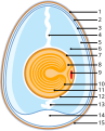 Vitelline membrane in a bird egg (7)
 -
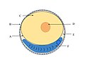
 Vitelline membrane in an amphibian egg (2)
 -
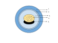
 Vitelline membrane in a fish egg (A)
 -
 Formation of Fertilization envelope from the Vitelline envelope
 -
 Parasitism and the Vitelline Membrane


.jpg)

_TEM.png)
See also[edit]
References[edit]
- ^ a b Damaziak, Krzysztof; Kieliszek, Marek; Bucław, Mateusz (30 January 2020). "Characterization of structure and protein of vitelline membranes of precocial (ring-necked pheasant, gray partridge) and superaltricial (cockatiel parrot, domestic pigeon) birds". PLOS ONE. 15 (1): e0228310. Bibcode:2020PLoSO..1528310D. doi:10.1371/journal.pone.0228310. ISSN 1932-6203. PMC 6992205. PMID 31999757.
- ^ a b Bellairs, Ruth; Osmond, Mark (2014). "Chapter 1 - The Hen's Egg and its Formation". Atlas of Chick Development (Third ed.). Academic Press. pp. 1–6. doi:10.1016/B978-0-12-384951-9.00001-0. ISBN 978-0-12-384951-9. Retrieved 23 October 2020.
- ^ a b Karlheinz, Mann Dr. (2008). "Proteomic analysis of the chicken egg vitelline membrane". Proteomics. 8 (11): 2322–2332. doi:10.1002/pmic.200800032. PMID 18452232. S2CID 206361990. Retrieved 2020-10-22.
- ^ Barresi, Michael J.F; Gilbert, Scott F. (July 2019). Vitelline envelope. Oxford University Press. pp. 220–221. ISBN 978-1605358222.
- ^ Tian, Jingdong; Gong, Hui; Thomsen, Gerald H.; Lennarz, William J. (1997). "Gamete Interactions in Xenopus laevis: Identification of Sperm Binding Glycoproteins in the Egg Vitelline Envelope". Journal of Cell Biology. 136 (5): 1099–1108. doi:10.1083/jcb.136.5.1099. PMC 2132474. PMID 9060474.
- ^ a b "vitelline membrane". Science Direct. Retrieved 2020-10-22.
- ^ a b c Lim, Whasun; Song, Gwonhwa (2015). "Differential expression of vitelline membrane outer layer protein 1: hormonal regulation of expression in the oviduct and in ovarian carcinomas from laying hens". Molecular and Cellular Endocrinology. 399: 250–258. doi:10.1016/j.mce.2014.10.015. PMID 25458700. S2CID 37646343. Retrieved 2020-10-22.
- ^ Monroy, Alberto (February 18, 2020). "Fertilization - Events of fertilization". Encyclopedia Britannica. Retrieved 2020-10-04.
- ^ "Egg | biology". Encyclopedia Britannica. January 30, 2019. Retrieved 2020-10-04.
- ^ Balinsky, Boris Ivan (September 23, 2011). "Animal development - Preparatory events". Encyclopedia Britannica. Retrieved 2020-10-04.
![]() This article incorporates text in the public domain from page 45 of the 20th edition of Gray's Anatomy (1918)
This article incorporates text in the public domain from page 45 of the 20th edition of Gray's Anatomy (1918)