Jump to content

File:MultiPhotonExcitation-Fig7-doi10.1186slash1475-925X-5-36.JPEG

Page contents not supported in other languages.
This is a file from the Wikimedia Commons
From Wikipedia, the free encyclopedia

Original file (2,992 × 2,696 pixels, file size: 1.93 MB, MIME type: image/jpeg)

Summary

Description
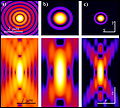
English: Original figure legend: Pointlike emitter optical response. From left to right: calculated x-y (above) and r-z (below) intensity distributions, in logarithmic scale, for a point like source imaged by means of wide-field, 2PE and confocal microscopy. Both 2PE and confocal shapes exhibit a better signal to noise ratio than widefield case. 2PE distribution is larger due the fact that a wavelength twice than in the wide-field and confocal case is responsible for the intensity distribution. Such intensity distributions are also known as point spread functions of the related microscopes. Optical conditions: excitation wavelengths are 488 nm and 900 nm for 1PE and 2PE, respectively; emission wavelength is 520 nm; numerical aperture is 1.3 for an oil immersion objective with oil refractive index value set at 1.515.
Deutsch: Original Abbildungslegende: Optische Antwort eines punktförmigen Emitters. Von links nach rechts: berechnete xy (oben) und rz (unten) Intensitätsverteilung in logarithmischer Skala für eine punktförmige Quelle aufgenommen in einem Weitfeld-, 2-Photonenanregungs- und Konfokal-Mikroskop. Sowohl die 2-Photonenanregungs, als auch die konfokalen Verteilungen zeigen ein besseres Signal-zu-Rausch-Verhältnis als im Fall des Weitfeldbildes. Das 2Photonenanregungsbild is weiter weil die benutzte Wellenlänge im Vergleich doppelt so groß ist. Diese Verteilungen sind auch als Punktspreizfunktionen ihrer Mikroskope bekannt. Optische Parameter: Anregungswellenlänger bei 488 nm für 1 Photonenanregung und 900 nm für 2 Photonenanregung; Emissionswellenlänge bei 520 nm und numerische Apertue 1.3 für ein Öl-Immersionsobjektiv mit einem angenommenen Brechungsindex für Öl bei 1.515.
Date
Source

Multi-photon excitation microscopy. BioMedical Engineering OnLine, 2006, 5:36.
DOI:10.1186/1475-925X-5-36.

http://www.biomedical-engineering-online.com/content/5/1/36
Author Alberto Diaspro, Paolo Bianchini, Giuseppe Vicidomini, Mario Faretta, Paola Ramoino and Cesare Usai
Permission
(Reusing this file)
w:en:Creative Commons
attribution
This file is licensed under the Creative Commons Attribution 2.0 Generic license.
You are free:
  • to share – to copy, distribute and transmit the work
  • to remix – to adapt the work
Under the following conditions:
  • attribution – You must give appropriate credit, provide a link to the license, and indicate if changes were made. You may do so in any reasonable manner, but not in any way that suggests the licensor endorses you or your use.

All images uploaded from this article about multi-photon and two-photon-microscopy:

Captions

Add a one-line explanation of what this file represents

Items portrayed in this file

depicts

6 June 2006

2,023,011 byte

2,696 pixel

2,992 pixel

image/jpeg

cc2098df1203db410625a405bf458cd58c5d2e3f

File history

Click on a date/time to view the file as it appeared at that time.

Date/TimeThumbnailDimensionsUserComment
current18:57, 23 December 2008Thumbnail for version as of 18:57, 23 December 20082,992 × 2,696 (1.93 MB)Dietzel65== Beschreibung == {{Information |Description={{en|1=Original figure legend: ''Pointlike emitter optical response. From left to right: calculated x-y (above) and r-z (below) intensity distributions, in logarithmic scale, for a point like source imaged by

Global file usage

The following other wikis use this file:

Metadata