
File:Phragmosome.png
Phragmosome.png (369 × 467 pixels, file size: 12 KB, MIME type: image/png)
| A vector version of this image is also available, and should be used in place of this raster image where the raster image contains information that could be stored more efficiently and/or accurately in the SVG format, as a vector graphic.
If its license requires the preservation of attribution or revision history, the raster version of this image should not be deleted, in order to maintain this information. For more information, see the documentation on MediaWiki's support of SVG images. |  |
Summary[edit]
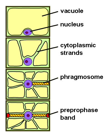
Phragmosome formation in a highly vacuolated plant cell. From top to bottom: 1) Interphase cell with large central vacuole. 2) Cytoplasmic strands starting to penetrate vacuole. 3) Nucleus migration into center and formation of the phragmosome. 4) Phragmosome formation completed and formation of preprophase band marking future cell division plane. When mitosis is completed, the new cell wall will form starting from the center along the plane occupied by the phragmosome.
References[edit]
- P.H. Raven, R.F. Evert, S.E. Eichhorn (2005): Biology of Plants, 7th Edition, W.H. Freeman and Company Publishers, New York, ISBN 0-7167-1007-2
Licensing[edit]
| I, the copyright holder of this work, hereby release it into the public domain. This applies worldwide. If this is not legally possible: |
File history
Click on a date/time to view the file as it appeared at that time.
| Date/Time | Thumbnail | Dimensions | User | Comment | |
|---|---|---|---|---|---|
| current | 04:55, 10 February 2007 | | 369 × 467 (12 KB) | Tameeria (talk | contribs) | Phragmosome formation in a highly vacuolated plant cell. From top to bottom: 1) Interphase cell with large central vacuole. 2) Cytoplasmic strands starting to penetrate vacuole. 3) Nucleus migration into center and formation of the phragmosome. 4) Phragmo |
You cannot overwrite this file.