
User:MiriamGonzalez14/Célula animal
An animal cell is aeukaryotic cell of which the different tissues of animals are composed.

1. Core.
1.1. Nuclear pore. 1.2. Chromatine. 1.3. Nuclear envelope.
1.4. Core. 1.5. Nucleolus.
2. Plasma membrane.
3. Golgi complex (vesicles, apparatus).
4. Ribosomes.
5. Rough endoplasmic reticulum.
6. Smooth endoplasmic reticulum.
7. Actin filaments.
8. Scourge.
9. Peroxisome.
10. Microtubule.
11. Lysosome.
12. Frees ribosomes.
13. Mitochondria.
14. Intermediate fibers.
15. Cytoplasm.
16. Secretory vesicle.
17. Centrosome (with two centrioles).
Structure[edit]
The large structure of animal cells can be divided in eight parts:
- The cellular envelope, constituted by the cellular membrane andplasma membrane
- The cytoplasm, in which cellular organelles are found: mitochondria, lysosomes,golgi apparatus,smooth endoplasmic reticulum, rough endoplasmic reticulum, centrioles, and ribosomes.
- The cellular nucleus, formed by the nuclear membrane that encompasses the nucleoplasm in which the chromatin and thenucleolus are located.
Cellular membrane, plasma membrane o plasmalema.[edit]
It is the extern limit of eukaryotic cells. It is a dynamic structure formed by two layers of phospholipids, in which molecules of cholesterol and proteins are embedded. The phospholipids have a hydrophilic head and two hydrophobic tails. The two layers of phospholipids are placed with the heads outward and the tails facing each other inward. That is, the hydrophilic groups are directed towards the aqueous phase, those of the outer layer of the membrane towards the extracellular liquid and those of the inner layer toward the cytoplasm. Its function is to delimit the cell and control what comes out and enters the cell.
Cytoplasm.[edit]
The cytoplasm is the part of a protoplasm that, in the eukaryotic cells, found between the cell nucleus and the plasma membrane.[1][2] It consists of a very fine colloidal emulsion of granular appearance, thecytosol or hialoplasma, and a variety of cellular organelles that perform different functions.
Its function is to house the cellular organelles and contribute to the movement of these. The cytosol is the seat of many of themetabolic processes that occur in cells.
The cytoplasm is divided in a external gelatinous region, close to the membrane, and involved in cell movement, which is called ectoplasm; and a more fluid internal part that is called the endoplasm and where most organelles are found.[3]
It is subdivided for a membrane network (smooth endoplasmic reticulum andrough endoplasmic reticulum) that serve as a work surface for many of its biochemical activities. In it are several nutrients that managed to cross the plasma membrane, thus reaching the organelles of the cell.
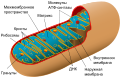
Mitochondria.[edit]
Small double membrane cell structure responsible for the conversion of nutrients into the energy-rich compound adenosine triphosphate (ATP), which acts as a cellular fuel. For this function they perform, called cellular respiration, it is said that mitochondria are the engine of the cell.
-
 Parte importante de la célula animal

Lysosome.[edit]
I take out a membrane that is found in cells with nucleus [EUCARIONTE] and contains hydrolytic enzymes that degrade complex molecules, such as [LEUKOCYTES] that destroy invaders and cell debris.
Golgi apparatus.[edit]
Differentiated part of the membrane system in the cellular interior, which is found in both animal and plant cells and has the function of modifying and distributing the proteins synthesized in the ribosomes of the granular or rough endoplasmic reticulum. These are transported in transition vesicles that fuse with the membrane of the Golgi cistern closest to the nucleus. Then, the proteins will be transferred through cisterns; finally, secretory vesicles are released containing the processed proteins throughout the entire apparatus. These vesicles will fuse with the plasma membrane, releasing their contents to the cellular exterior. During the transport through the different tanks of the Golgi, the proteins are modified, since they are added glucides or fatty acids.
Endoplasmic reticulum.[edit]
The reticulum endoplasmic is an complex sistem of membranes arranged in the form of flattened sacs and tubules that are interconnected with each other sharing the same internal space. Their membranes are continued with those of the nuclear envelope and can extend to the vicinity of the plasma membrane, reaching less than half of the membranes of a cell. Because the fatty acids that compose them tend to be shorter, they are thinner than the others [4]
The reticulum organizes its membranes in regions or domains that perform different functions. The two domains that are easiest to distinguish are the rough endoplasmic reticulum, with its membranes forming more or less straight tubules, sometimes flattened cisterns, and with numerous associated ribosomes, and the smooth endoplasmic reticulum, without associated ribosomes and with membranes organized to form very tubular curved and irregular[4]
The outer membrane of the nuclear envelope can be considered part of the endoplasmic reticulum since it is a physical continuation of it and ribosomes associated with it can be observed carrying out the translation. The rough and smooth endoplasmic reticulum usually occupy different cell spaces as in hepatocytes, in neurons and in cells that synthesize steroids. However, in some regions of the reticulum there is no clear segregation between both domains and there are areas of membrane with ribosomes mixed with others without ribosomes. The spatial arrangement of the endoplasmic reticulum in animal cells depends on their interactions with microtubules, while in plants it is theactin filaments that are responsible.[4]
Rough Reticulum Endoplasmic or RRE.[edit]
The rough endoplasmic reticulum is present in all eukaryotic cells (nonexistent inprokaryotes) and predominates in those that manufacture large amounts ofproteins for export. It continues with the outer membrane of the nuclear envelope, which also has attached ribosomes. Its outer surface is covered with ribosomes, where protein synthesis occurs. It transports the proteins produced in the ribosomes to the cellular regions where they are necessary or to the Golgi apparatus, from where they can be exported abroad.
Smooth Reticulum Endoplasmic or SRE.[edit]
The smooth endoplasmic reticulum performs several functions. It intervenes in the synthesis of almost all the lipids that form the cell membrane and the other membranes that surround the other cellular structures, such as mitochondria. Cells specialized in lipid metabolism, such as liver cells, tend to have more smooth ER. Smooth ER also intervenes in the absorption and release of calcium to mediate some types of cellular activity. In skeletal muscle cells, for example, the release of calcium by the ER activates muscle contraction.
Centriole.[edit]

A centriole or centriole is an organelle with a cylindrical structure, consisting of 9 triplets of microtubules, which is part of the cytoskeleton. A pair of centrioles positioned perpendicularly to each other and located inside a cell is called diplosome. When the diplosome is surrounded by pericentriolar material (a dense protein mass), it is called a centrosome or microtubule organizing center (COMT), which is characteristic of animal cells.
Centrioles allow the polymerization of microtubules of tubulin dimers, which are part of the cytoskeleton and which are irradiated therefrom by a star-like arrangement called a mitotic spindle.
In addition, they intervene in cell division, contribute to the maintenance of cell shape, transport organelles and particles inside the cell, form structural elements such as the mitotic spindle and form the cytoskeletal axis in eukaryotic cilia and flagella, as well as the of the basal corpuscles.
Core.[edit]
It is the most conspicuous organ in almost all animal and plant cells, is surrounded by a characteristic membrane, is spherical and measures about 5.2 μm in diameter. Within the nucleus, the DNA and protein molecules are organized into chromosomes that usually appear arranged in identical pairs. One of the main characteristics is that it is of small body of spheroid or oval aspect, it is mostly in the center of the nucleus and that some can be located in the periphery of the cell.
Nucleoplasm.[edit]
The nucleus of the eukaryotic cells is a discrete structure that contains the ribosomes, recipients of the genetic endowment of the cell. It is separated from the rest of the cell by a double-layer nuclear membrane and contains a material called nucleoplasm. The nuclear membrane is perforated by pores that allow the exchange of cellular material between nucleoplasm and cytoplasm.
Cromatine.[edit]
The cromatine is the set of DNA, histone proteins and non-histone proteins, found in the nucleus of eukaryotic cells and constituting the genome of these cells.
The basic units of chromatin are nucleosomes. These are formed by approximately 146 base pairs in length (the number depends on the organism), associated with a specific complex of 8 nucleosomal histones (histone octamer). Each particle has a disk shape, with a diameter of 11 nm and contains two copies of each of the 4 histones H3, H4, H2A and H2B. This octamer forms a protein nucleus, around which the DNA helix is wound (approximately 1.8 turns). Between each of the associations of DNA and histones there is a free DNA called DNA spacer, variable length between 0 and 80 nucleotide pairs that guarantees flexibility to the chromatin fiber. This type of organization allows a first step of compaction of the genetic material, and gives rise to a structure similar to a "necklace of beads".
Subsequently, a second level of organization of higher order is the "30nm fiber", composed of groups of nucleosomes packed over each other, adopting regular arrangements thanks to the action of histone H1.
Finally, the increase in DNA packing continues until we get the chromosomes we observed in the metaphase, which is the maximum level of DNA condensation.
Nucléolus.[edit]
The nucleolus is a region of the nucleus that is considered a supra-macromolecular structure, which does not have a membrane that limits it. The main function of the nucleolus is thetranscription of ribosomal ribonucleic acid (rRNA) by polymerase I, and the subsequent processing and assembly of the pre-components that will form the ribosomes. The biogenesis of the ribosome is a very dynamic nucleolar process, involving: the synthesis and maturation of rRNA, its transient interactions with non-ribosomal proteins and ribonucleoproteins and, also, the assembly with ribosomal proteins.[5]
In addition, the nucleolus has roles in other cellular functions such as regulation of the cell cycle, cellular stress responses, telomerase activity and aging.
These facts show the multifunctional nature of the nucleolus, which is reflected in the complexity of its protein and RNA composition, and is also reflected in the dynamic changes that its molecular composition presents in response to variable cellular conditions.[6]
Comparison with other eukaryotic cells.[edit]
The animal cell differs from other eukaryotic cells, mainly plant cells, in that it lacks a cell wall andchloroplasts, and in that it possesses smaller vacuoles. Due to the absence of a rigidcell wall, animal cells can take a variety of forms, and can even surround and engulf other structures, such as phagocytic cells.
| Tipic animal cell | Tipic vegetable cell | |
|---|---|---|
| Basic structures | ||
| Organules |
|
|
| Additional structures |
|
| Célula animal típica | Célula vegetal típica |
|---|---|
|
3. Ribosomas |
h. Mitocondria
k. r. Vesículas |


Referencias[edit]
- ^ "citoplasma". WordReference. 2005. Retrieved 26 October 2007.
- ^ "Definición de citoplasma". definición.org. Retrieved 26 October 2007.
- ^ "Letra E". Diccionario Ecológico. Ambiente Ecológico. Retrieved 26 October 2007. ISSN 1668-3358
- ^ a b c "Retículo endoplasmático". Atlas de Histología Vegetal y Animal. Facultad de Biología de la Universidad de Vigo, Galicia: Departamento de Biología Funcional y Ciencias de la Salud. 13 September 2013. Retrieved 14 February 2014.
{{cite web}}: External link in|work= - ^ Ivan Raška, Karel Koberna, Jan Malínský, Helena Fidlerová, Martin Mašata 2004; El nucléolo y la transcripción de genes ribosomales, Biology of the Cell 96 (2004) 579-594.
- ^ Boisvert, Francois-Michel (2007). "The Multifunctional nucleolus". Nature Reviews Molecular Cell Biology. Vol. 8 (July 2007).
Enlaces externos[edit]
Diagrams of cells & cell ultrastructure [[Category:Cells]] [[Category:Animal physiology]]