
User:Thecurran91/methyl violet

| |
| Names | |
|---|---|
| IUPAC name
Methyl violet 2B
| |
| Identifiers | |
3D model (JSmol)
|
|
| UNII | |
| |
| Properties | |
| C24H28N3Cl | |
| Molar mass | 393.958 g/mol (6B) |
| Melting point | 137 °C (279 °F) |
Except where otherwise noted, data are given for materials in their standard state (at 25 °C [77 °F], 100 kPa).
| |
Methyl violet is the name given to a group of similar chemicals used as pH indicators and dyes. Methyl violets are mixtures of tetramethyl, pentamethyl and hexamethyl pararosanilins. By blending the different versions, the dyemaker can create different shades of violet in the final dye. The more methylated the compound (the more methyl groups attached), the deeper blue the final color will be:
- Tetramethyl (four methyls) is known as methyl violet 2B, and this specific chemical finds uses in chemistry and medicine.
- Pentamethyl (five methyls) is known as methyl violet 6B, and is darker (in dye form) than 2B.
- Hexamethyl (six methyls) is known as methyl violet 10B, or specifically as crystal violet. This is much darker than 2B, and often darker than 6B.
In pure form, the tetramethyl appears as lustrous blue-green crystals that melt at 137°C (279°F).
The main use of methyl violet (by sheer volume used worldwide) is to dye textiles purple and give deep violet colors in paints and printing ink.
Methyl violet 2B (simply called methyl violet) is used in chemistry as a pH indicator to test pH ranges from 0 to 1.6. At the acid end of its measuring range, it takes on a yellow color. At the alkaline end, it becomes bluish-violet. Methyl violet can be supplied as premade pH testing paper, or it can be supplied as pure crystals and dissolved in the sample being checked.
In medicine, Methyl Violet 10B is known as Gentian violet and is the active ingredient in Gram's stain, used to classify bacteria. Gentian violet destroys cells, and is used as a moderate-strength external disinfectant. Gentian violet is very poisonous to most animals, dogs and cats included — it should never be used as a disinfectant for animals' skin. As Methyl Violet inhibits the the growth of most Gram positive organisms other than streptococci, it can be used in conjunction with nalidixic acid (which inhibits or kills gram negative bacteria) in order to isolate streptococci in the diagnoses of streptococcal infections.
Methyl violet has also the ability of binding DNA. Therefore, in biomedical sciences, it is used for cell viability assays. The binding to DNA can also cause disruption in DNA replication process, which can lead to mutations and cancers.
Methyl violets are soluble in water, ethanol, diethylene glycol, and dipropylene glycol. Specifically, methyl violet 6B is 2.93% soluble in water and 15.21% soluble in ethanol.
Methyl violet should not be confused with methyl blue or methylene blue, two other stains.
-
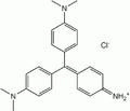 Methyl Violet 2B
 -
 Methyl Violet 6B
 -
 Methyl Violet 10B


Degradation[edit]
Methyl violet is a mutagen and mitotic poison, therefore concerns exist regarding the ecological impact of the release of methyl violet into the environment. Methyl violet has been used in vast quantities for textile and paper dyeing, and 15% of such dyes produced worldwide are released to environment in wastewater. Numerous methods have been developed to treat methyl violet pollution. The three most prominent are chemical bleaching, biodegradation, and photo degradation.
Chemical bleaching[edit]
Chemical bleaching is achieved by oxidation or reduction. Oxidation either destroys the dye completely or causes a change in the bonding of the chromophore[citation needed]. Two examples of dye oxidants are sodium hypochlorite (NaClO, common bleach) and hydrogen peroxide). NaClO produces hypochlorous acid (HClO), hypochlorite ions (ClO-) and chlorine, which are all in equilibrium.
- NaOCl + H2O ⇌ Na+ + OH− + H+ + OCl−

When any one of these compounds come in contact with the amine groups of the dye, they hydrolyze and degrade them. Methyl violet has three amine groups, and when one of more of these groups are hydrolyzed the molecule must rearrange itself to form a more stable compound. This breaks the chromophore bonds, meaning the molecule no longer absorbs light.
Hydrogen peroxide breaks the dye's bonds by forming radical species in the presences of light. These oxidize the dye by adding a oxygen atoms on to the nitrogen in the amine group.
The reduction of methyl violet mostly occurs in microorganisms but it can be attained chemically using sodium dithionite and sodium hydrosulfide[citation needed].
Biodegradation[edit]
Biodegradation is the most interesting and most investigated method of dye degradation. This method is suitable because biodegradation could occur in large sewage plants with specialized microorganisms- which is highly cost effective. Certain animals and plants can degrade this dye, as well as microorganisms[citation needed], but the microorganisms are the most practical solution.
Two microorganisms that have been studied in depth are the White Rot Fungus and the bacterium Nocardia Corallina. In particular, the White Rot Fungus degraded a 12.3 µM methyl violet solution to 35% of the initial concentration in 6 hours. In 12 hours only 1% remained and after 72 hours the dye was deemed to be completely degraded [citation needed].
Nocardia Carollina's growth was inhibited by the toxic dye at the start of an incubation, but was able to degrade dyes with a concentration of under 5 µmol cm-3. The bacteria were completely inhibited with concentrations higher than 7 µmol cm-3[citation needed].


Photo degradation[edit]
Light alone is not enough to cause major degradation of methyl violet[citation needed]. However, with the addition of large band-gap semiconductors, TiO2 or ZnO, the photodecomposition speeds up.
The mechanism behind the TiO2 catalysis is that is causes the production of oxygen free radicals, which break up the dye molecule. The rate of degradation can be increased by adding oxidisers or radical-forming molecules such as hydrogen peroxide, or Ag+ ions.

Other methods[edit]
Many others methods have been developed to treat the contamination of dyes in a solution such as:
Electrochemical Degradation[edit]
This is accomplished by running DC current through the dye solution to break the dye apart. This works well with dyes that are molecularly simple, but is ineffective against very complex dyes. This methods work very well when it is used to decomposed methyl violet. This method can be further improved by the addition of a redox mediator such as Co+2/+3 .
Ion Exchange Membrane[edit]
In this method a membrane is used to separate the cation of the dye from the solution. The experimental results indicates that the addition of an organic solvent containing ions (1M NaCl and 60% CH3OH) increases the separation of the cation from solution to 100%.
Laser Degradation[edit]
It was found that Kr-2 excited with a 530 nm laser allowed for electron transfer from the triplets state to the Cationic dye methyl violet. The addition of this electron to the cation forces the molecule to rearrange.
Absorption[edit]
Absorbance of the dye from solution has been observed using solid Porous material such as: Pumice powder Porous silicon additive Porous glass Activated charcoal Micro tubes Ceramics
Many methods are available for dye removal from waste water but most are expensive, impractical or they just allow for the polluted dye to leave from one source to the next. In current removal techniques many of the procedure listed are used in combinatioghjhgjghjn to degrade the dye in the waste water.
See also[edit]
- Potassium ferrocyanide
- Potassium ferricyanide
- Methylene blue
- Methyl blue
- Egyptian Blue
- Han Purple
- Gentian violet
- Fluorescein
References[edit]
- [*Kristallviolett – ein pH-Indikator (in German)
- Mechanism of action of sodium hypochlorite [1]
- Hydrogen Peroxide[ [2]
- XP-Chloro Degradation Malachite green US Patent 2755202)
- Senthilkumaar S., and Porkodi.(2005). Heterogeneous Photocatalytic Decomposition of Crystal Violet in UV-illuminated Sol Gel
- Derived Nanocrystalline TiO2 Suspension. Journal of Colloid and Interface Science. 288(1):184-189.
- Bumpus J.A. and Brook B. J.(1988). Biodegradation of Crystal Violet by the White Rot Fungus Phanerochaete chrysosporium. Applied and Environmental Microbiology.54(5):1143-1150.
- Pizzolato T. M. et al.(2002). Colour Removal with NaClO of Dye Wastewater From Agate-processing plant in Rio Grande do Sul, Brazil. International journal of Mineral Processing.65 (1-4):203-211.
- Bhasikuttan C.A. et al.(2008).Photo ionization of Crystal Violet in Aqueous Solution. Photochemistry and Photobiology.62(2):245-250.
- Yatome C. et al.(1993).Degradation of Crystal Violet by Nocardia corallina. Applied Microbiology and Biotechnology.38-565-569.
- Bhasikuttan A.C. et al.(1995). Oxidation of Chrystal Violet and Malachite green in aqueous solutions- a kinetic spectrophotometric study. Journal of Photochemistry and photobiology A:Chemistry. 90(2-3):177-182.
- Sahoo, A.K.G. and Pal, A.(2005. Photocalalytic Degradation of Cystal violet(C.I. Violet) on Dilver Ion Doped TiO2. Dyes and Pigments. 66(3):189-196.
- Saroman , M. A. et al.(2004). Electrochemical Decolonization of Structurally Different Dyes. Chemosphere. 57(3):233-239.
- Wu, J. et al. (2008). Removal of Catonic Dye Methyl Violet 2B From Water by Cation Exchange Membrances. Journal of Membrance Science.309(1-2):239-245.