User:Yoshita S/sandbox
GUNTHER DISEASE
Synonyms of Gunther disease[edit]
- CEP(congenital erythropoietic porphyria)
- Uroporphyrinogen III synthase deficiency
- UROS deficiency
Introduction[edit]

Congenital erythropoietic porphyria (CEP) is a very rare inherited metabolic disorder resulting from the deficient function of the enzyme uroporphyrinogen lll cosynthase (UROS), the fourth enzyme in the heme biosynthetic pathway. Due to the impaired function of this enzyme, excessive amounts of particular porphyrins accumulate, particularly in the bone marrow, plasma, red blood cells, urine, teeth, and bones. The major symptom of this disorder is hypersensitivity of the skin to sunlight and some types of artificial light, such as fluorescent lights (photosensitivity). After exposure to light, the photo-activated porphyrins in the skin cause bullae (blistering) and the fluid-filled sacs rupture, and the lesions often get infected. These infected lesions can lead to scarring, bone loss, and deformities. The hands, arms, and face are the most commonly affected areas. CEP is inherited as an autosomal recessive genetic disorder. Typically, there is no family history of the disease. Neither parent has symptoms of CEP, but each carries a defective gene that they can pass to their children. Affected offspring have two copies of the defective gene, one inherited from each parent.As is characteristic of the erythropoietic porphyrias, symptoms begin during infancy. Sometimes CEP is recognized as a cause of anemia in a fetus before birth. In less severe cases symptoms may begin during adult life.
Signs & Symptoms[edit]

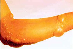
The most common symptom of CEP is hypersensitivity of the skin to sunlight and some types of artificial light (photosensitivity), with blistering of the skin occurring after exposure. Affected individuals may also exhibit abnormal accumulations of body fluid under affected areas (edema) and/or persistent redness or inflammation of the skin (erythema). Affected areas of the skin may develop sac-like lesions (vesicles or bullae), scar, and/or become discolored (hyperpigmentation) if exposure to sunlight is prolonged. These affected areas of skin may become abnormally thick. In addition, in some cases, affected individuals may also exhibit malformations of the fingers and nails. The severity and degree of photosensitivity differ depending on the severity of the patient’s gene lesions which correlate with the deficient enzyme activity. Photosensitivity is seen from birth; however, in some cases, it may not occur until childhood, adolescence or adulthood. Patients also have erythrodontia, brownish discolored teeth, which fluoresce under ultraviolet light.
In more severe cases, other symptoms can include a low level of red blood cells (anemia), enlargement of the spleen, and increased hair growth (hypertrichosis). The anemia can be severe and such patients require periodic transformations to maintain sufficient red blood cells. In severely affected patients, anemia may be present in the fetus. Ocular problems also can occur including corneal scarring, eye inflammation, and infections.
Symptoms usually start in infancy or childhood and the diagnosis in most patients is suggested by the reddish color of the urine which stains the diapers. The diagnosis is made by finding increased levels of specific porphyrins in the urine. Diagnostic confirmation is made by measuring the specific (UROS) enzyme activity and/or by identifying the specific lesion(s) in the UROS gene which is/are responsible for the impaired enzyme.
Causes[edit]

Congenital erythropoietic porphyria is inherited as an autosomal recessive genetic condition. Recessive genetic disorders occur when an individual inherits two copies of an abnormal gene for the same trait, one from each parent. If an individual receives one normal gene and one gene for the disease, the person will be a carrier for the disease, and usually will not show symptoms. The risk for two carrier parents to both pass the defective gene and have an affected child is 25% with each pregnancy. The risk to have a child who is a carrier like the parents is 50% with each pregnancy. The chance for a child to receive normal genes from both parents and be genetically normal for that particular trait is 25%. The risk is the same for males and females.
Parents who are close relatives (consanguineous) have a higher chance than unrelated parents to both carry the same abnormal gene, which increases the risk to have children with a recessive genetic disorder. Porphyria disorders affect the production of functional haem molecules in haemoglobin. The haem component is composed of a porphyrin ring complex and iron. Porphyria affects the production of a functional porphyrin complex through a genetic mutation at any one of the many enzymatic steps involved in its production. While most haem is in the blood associated with haemoglobin, haem is also required for in several other tissues, including the liver. Porphyrias can affect either the skin (cutaneous porphyria) or the nervous system (acute porphyria).
Mutations in the UROS gene cause CEP. The symptoms of CEP develop due to excessive levels of the specific porphyrins that accumulate in tissues of the body as a result of the markedly impaired function of the UROS enzyme.
Affected Populations[edit]
CEP is a very rare genetic disorder that affects males and females in equal numbers. Over 200 cases have been reported worldwide.

Diagnosis[edit]
The diagnosis of CEP may be suspected when the reddish-colored urine is noted at birth or later in life. This finding, or the occurrence of skin blisters on sun or light exposure, should lead to a thorough clinical evaluation and specialized laboratory tests. The diagnosis can be made by testing the urine for increased levels of specific porphyrins. Diagnostic confirmation requires the demonstration of the specific UROS enzyme deficiency and/or the lesion(s) in the UROS gene.
Prenatal and preimplantation genetic diagnoses are available for subsequent pregnancies in CEP families.
Standard Therapies[edit]
Treatment[edit]
- Avoidance of sunlight is essential to prevent the skin lesions in individuals with CEP.
- he use of topical sunscreens, protective clothing, long sleeves, hats, gloves, and sunglasses are strongly recommended.

Bone marrow transplant - Individuals with CEP will benefit from window tinting or using vinyls or films to cover the windows in their car or house.
- In addition to protection from sunlight, the anemia should be treated, if present. Chronic transfusions have been useful in decreasing the bone marrow production of the phototoxic porphyrins.
- bone marrow transplantation has cured patients with CEP, but is accompanied by specific risks of complications and demise.
Investigational Therapies[edit]
Porphyrin production in the bone marrow can be reduced by red blood cell (erythrocyte) transfusions but must be used with caution due to complications associated with chronic transfusion therapy.
Successful bone marrow transplantation has been curative for patients now over 10 years post-transplantation. Hematopoietic stem cell cord blood transplantation has also been successful.
The Rare Diseases Clinical Research Network (RDCRN), funded by the National Institute of Health (NIH) and the Office for Rare Diseases Research (ORDR), is sponsoring The Porphyrias Consortium, which will focus on the inborn errors of heme biosynthesis, the Porphyrias. It is bringing together senior porphyria experts at six academic institutions, the American Porphyria Foundation (APF), and industry to carry out clinical studies and clinical trials to accelerate the development of improved diagnosis and treatment for the patients with these rare diseases
Case Report[edit]
A 15-year-old female patient came to our outpatient department with a concern for her discolored teeth and slowly growing swelling around the right side of the jaw. Mother had noticed that soon after birth, her daughter began to get sunburned easily, with noticeable blister formations on exposed areas which required sun protection. When she was about 1 year old, her urine color changed gradually from red-purple to dark-purple. Her teeth became brownish-purple (erythrodontia) while she was in elementary school, and hypertrichosis developed with irregular hypo and hyper-pigmentation over unprotected skin areas on reaching puberty. Since her secondary school years, she suffered from frequent malaise and anemia with hemoglobin fluctuating between 8 and 11 g%. The patient had a healthy younger brother, no family history of porphyria and no history of consanguinity.
Physical examination revealed extensive brownish-purple blisters and scarring over the face, lips, forehead, ears, mutilation of toe fingers, excessive facial hair, and hypertrichosis of the ears. A diffuse, nontender, firm extra-oral swelling over the right body of mandible was also evident. Intra-oral examination revealed marked cervical generalized purplish pigmentation of the teeth [Figure 4] which fluoresced under ultraviolet light. Porphyria was diagnosed for the cutaneous condition, with consideration of CEP as the possible subtype. Displacement of the mandibular right lateral incisor, first and second premolars, and a missing canine along with a smooth surfaced diffuse round to ovoid swelling with normal appearing overlying mucosa was seen involving the body of mandible, obliterating the mandibular vestibule from lateral incisor to the second premolar. Lymphadenopathy was not evident. Dental findings suggested an odontogenic pathology, and a differential diagnosis of dentigerous cyst, keratocystic odontogenic tumor, calcifying odontogenic cyst and AOT were considered.
Orthopantomograph revealed a unilocular radiolucency with a well-defined cortex associated with an impacted mandibular right canine causing displacement and root divergence, but no root resorption of the adjacent lateral incisor and premolars. There was some amount of underlying alveolar bone erosion and thinning of the inferior cortex. Bilateral carious involvement of mandibular first molars with periapical infection was also seen. Based on the radiographic findings a differential diagnosis of AOT or keratocystic odontogenic tumor was reached.
The preceding history, physical examination, and laboratory findings were in confirmation with the clinical impression of CEP. Patient was advised bone marrow transplantation but refused due to fear of possible complications and accepted avoiding sunlight as the only treatment, as most kinds of sunscreen were ineffective. Patient was advised to wear clothing that would cover the exposed skin parts, in an event of venturing outside, so as to minimize the cutaneous photosensitivity.
FAQs[edit]
1. What is congenital erythropoietic porphyria?
Congenital erythropoietic porphyria (CEP), also called Günther’s disease after the doctor who first described it, is the rarest of the porphyrias. It is estimated that about 1 in every 2 – 3 million people are affected by CEP, which affects males and females equally, and occurs in all ethnic groups.
2. What causes congenital erythropoietic porphyria?
In CEP, there are high levels of a porphyrin called porphyrinogen in the bone marrow, blood and urine, which cause the symptoms and signs.
3. Is congenital erythropoietic porphyria hereditary?
Yes. The parents of someone with CEP have no symptoms of the condition themselves (and are called carriers of the condition), but each of them has a mutation in one of their genes. There is a 1 in 4 risk that each child born to 2 carriers will inherit the abnormal gene from both parents and thus develop the condition. This form of inheritance is called autosomal recessive.
4. What are the symptoms of congenital erythropoietic porphyria?
Individuals with CEP may not have all of the problems described in this leaflet as the severity of the condition varies. Usually, the disease shows itself soon after birth or in early childhood, but sometimes onset of disease is delayed until the teenage years or early adulthood.
    * Red urine is usually the first sign noticed in newborn babies with CEP. The intensity of the redness can vary from day to day.
    * The skin is very sensitive to light, especially direct sunlight, which may cause blisters or ulcers, which heal to leave scars. This most commonly happens at sun-exposed sites, for example the backs of the hands, the face, ears and scalp.
    * The eyes may also be sensitive to bright sunlight or artificial light, which can cause ulcers and scarring. With time, some patients lose their eyelashes, making their eyes easily irritated by small particles of dust    and fibre.
    * The skin may take longer to heal after injury or blistering, and become infected.
    * Anaemia, which varies in severity, is common in CEP. Anaemia develops because porphyrin damages red blood cells, and causes tiredness, shortness of breath following minimal exertion, and paleness.
    * The spleen, which removes the damaged red blood cells, can gradually become bigger and cause worsening of the anaemia and a reduction in the number of platelets (the blood cells that help to form blood clots to stop bleeding) and white cells (the blood cells that fight infections) in the blood. This can lead to an increased risk of bleeding (such as repeated nose bleeds) and infections.
    * CEP can occasionally cause thinning of the bones (osteoporosis), which can lead to bone fractures following minor injury.
5. Can CEP be cured?
Currently, the only available cure for CEP is a bone marrow transplant (BMT). This involves transplanting healthy bone marrow from another person (the donor) to the person with CEP (the recipient). Following successful BMT, the symptoms of CEP such as photosensitivity and anaemia will improve. However, the scarring from previous damage to the skin is permanent.
References[edit]
http://www.porphyriafoundation.com/about-porphyria/types-of-porphyria/CEP

