
Wikipedia:Featured picture candidates/Plant cell structure
Plant cell structure[edit]


Created by Commons:User:LadyofHats over on Commons. Incredibly informative to all articles that it appears in.
- Nominate and support. - Kilo-Lima|(talk) 13:40, 26 May 2006 (UTC)
- Support Text book quality. An SVG would be even better.-Ravedave 15:11, 26 May 2006 (UTC)
- Should be SVG'd, and I'm not sure about the green border and the caption. ed g2s • talk 16:43, 26 May 2006 (UTC)
- Support. Can PNGs be converted to SVGs? JQF 17:31, 26 May 2006 (UTC)
- Support with or without caption. --BRIAN0918 20:15, 26 May 2006 (UTC)
- Support informative and very nice diagram. Pegasus1138Talk | Contribs | Email ---- 20:34, 26 May 2006 (UTC)
- Support a great diagram of a plant cell. Anonymous_anonymous_Have a Nice Day 20:43, 26 May 2006 (UTC)
- Oppose. The green framing is not needed, and one of the callouts is half in the green. And yes, an SVG would be ideal.--ragesoss 20:53, 26 May 2006 (UTC)
- I have fixed the framing though due to the way the callout is done it is extremely hard to move it around without botching either it or the background so for the moment I have left it as is and will be working a a version with that change later. Pegasus1138Talk | Contribs | Email ---- 21:46, 26 May 2006 (UTC)
- Extremely difficult? I just copied and pasted letters from other words into place. --BRIAN0918 22:29, 26 May 2006 (UTC)
- For the membraneous vesticles? it contains letters not contained in any of the other words and the ones in that section are hard to make use of without overly fading / destroying due to the green background. Pegasus1138Talk | Contribs | Email ---- 05:49, 27 May 2006 (UTC)
- weak oppose for borderless version; there is too much white space around the edge, and (in both versions) the dark green shading doesn't line up with the black outlines, especially on the lower right. If there was color spilling over all around it, in natural-looking ways, this might be stylish; as it is, it looks sloppy. Regarding the callout, I was talking about filamentous cytoskeleton; the one that touches the actual diagram is not a problem, in my opinion.--ragesoss 00:45, 28 May 2006 (UTC)
- For the membraneous vesticles? it contains letters not contained in any of the other words and the ones in that section are hard to make use of without overly fading / destroying due to the green background. Pegasus1138Talk | Contribs | Email ---- 05:49, 27 May 2006 (UTC)
- Extremely difficult? I just copied and pasted letters from other words into place. --BRIAN0918 22:29, 26 May 2006 (UTC)
- Oppose PNG, but I would support SVG. There is absolutely no reason to upload this kind of illustration as PNG other than not knowing how to save it as SVG. What application was used to create the picture? It should be no problem to generate an SVG version from the original (not from the PNG though). --Dschwen 10:44, 27 May 2006 (UTC)
- Support version without green border. There's absolutely no reason to oppose an image just because it's not in an SVG format. That should only be an issue if it affects the quality of the image, which this doesn't. - Mgm|(talk) 14:09, 27 May 2006 (UTC)
- Actually it does affect the quality of this image. If scaled to higher resolutions (e.g. for print) this PNG will not look sharp, whereas SVGs scale infinitely well. Redquark 16:54, 27 May 2006 (UTC)
- Support version without green border. Would prefer SVG version though. --BillC 20:12, 27 May 2006 (UTC)
- Comment - Interestingly, the analogous animal cell diagram is an SVG, as are LadyofHats' other recent contributions. I think we should put this nomination on hold until she has a chance to respond to the request for an improved version. We all agree this is a great diagram, but most people also think it could be better by being an SVG (in which case we could also tweak it further without much trouble). Let's not settle settle for less than the best.--ragesoss 01:03, 28 May 2006 (UTC)
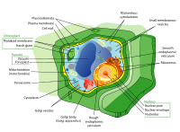
Comment/Oppose- While this illustration does some things well there are a few iffy things on the science front.
- The leukoplast (most commonly spelled leucoplast) is a non-pigemented plastid (i.e. chloroplast) and typically only occurs in non-photosynthetic tissues - which is slightly problematic since this cell also has chloroplasts (and it should since they are a defining character of a "plant cell") - and should probably be removed for simplicity and accuracy.
- The membrane around the vacuole is called the tonoplast - this should be added, the vacuole also takes up a lot of the cell - which is not really reflected here. The "wheel of cheese with a wegde removed" seems to be the best way to demonstrate with spatial relatioship between organelles in cells.
- The spatial relationship of the rough and sooth ER and the golgi is kind of weird - and might be better illustrated more like this - the smooth ER in the FPC are disconnected in this FPC image - when they are in fact continuous - and I think the "small membranous vesicles" shouldn't be there (vesicles are small membrane bound compartments that move stuff between parts of the cell, especially in the ER - they are not big wormy things). It should probably also be shown that the rough ER is rough because of the association of ribosomes. The golgi is usually just referred to ad a Golgi body.
- The filamentous cytoskeleton should probably be a different colour so it's not confused with the ER.
- Why does cytosol label point to a red circle? when cytosol fills the whole cell?
- For clarity things that are a part of a superstructure should be labelled together like this encarta diagram - which is not accurate but has nice clear labels.
- --Peta 03:58, 29 May 2006 (UTC)
Oppose. Shouldn't be featured until Peta's objections are addressed, and no edits seem to be forthcoming. Although there are more support votes than opposes, I think an FAC-style "one actionable objection is enough to sink the nom" standard should be applied in this case. Redquark 19:46, 5 June 2006 (UTC)- Support updated version. Redquark 23:01, 6 June 2006 (UTC)
last SVG version

The plasma membrane is a layer that is in between the cytoplasm and the cell wall.
- Here is the last SVG version with all the changes you have asked.
- to answer to your questions:
- the reason why the original is a PNG, is becouse to the time i was new on wikipedia and someone had told me that all diagrams should be in this format. later on someone else told me the same about SVG. and since this last one seems a much more practical format i started to work on it NOTE: there is a proplem with SVG format on wikipedia. the image will must probably do not show on galleries, and depending on the size of the thumb it will show or not on the article. i was hoping this would be fixed soon, meanwhile one can try to fix it by making a thumb instead of a gallery and changing the size of the thumb in one or two pixels.
- PNGs can not be converted to SVGs, since one is a pixel based format and the other is a vector based format.
- i actually liked the green border and to have the title inside the image becouse it lets the image stand alone without the need of the article; this allows the image to be used inside and outside wikipedia. i had in mind that the image was being released as public domain and that wikipedia images apear in many other websites, so i wanted the image to explain things by itself . this is also a reason why i type the names of the elements and not just numbers as many other people do. Even then i was told already several times that borders are not desired in wiki proyects. so i do not do them anymore :P.
- on the elements names and why they apear, i based myself in the examples i had on hand. both in form , position and name. i know is posible there are mistakes and i am always open to fix them. you just had to tell me :)
- Thankyou all for taking so much time for one of my images. if you need anything, just leave me a mesage LadyofHats 09:50, 6 June 2006 (UTC)
- Support updated version, although I think the small membrane bound vesicles are an unnecessary and potentially confusing addition.--Peta 11:56, 6 June 2006 (UTC)
Promoted Image:Plant cell structure-en.svg Raven4x4x 09:47, 7 June 2006 (UTC)