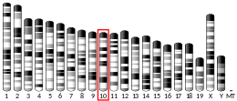
CD63
CD63 antigen is a protein that, in humans, is encoded by the CD63 gene.[5] CD63 is mainly associated with membranes of intracellular vesicles, although cell surface expression may be induced.
Function
[edit]The protein encoded by this gene is a member of the transmembrane 4 superfamily, also known as the tetraspanin family. Most of these members are cell-surface proteins that are characterized by the presence of four hydrophobic domains. The proteins mediate signal transduction events that play a role in the regulation of cell development, activation, growth, and motility. This encoded protein is a cell surface glycoprotein that is known to complex with integrins. It may function as a blood platelet activation marker. Deficiency of this protein is associated with Hermansky-Pudlak Syndrome . Also this gene has been associated with tumor progression. The use of alternate polyadenylation sites has been found for this gene. Alternative splicing results in multiple transcript variants encoding different proteins.[5]
Allergy diagnosis
[edit]CD63 is a good marker for flow cytometric quantification of in vitro activated basophils for diagnosis of IgE-mediated allergy. The test is commonly designated as basophil activation test (BAT).
Research
[edit]Initially, deletion and point mutants were used to investigate the role of the C-terminus, which contains a putative lysosomal-targeting/internalisation motif (GYEVM). C-terminal mutants showed increased surface expression and decreased intracellular localisation relative to CD63Wt. Antibody induced internalisation was reduced in C-terminal deletion mutants and abolished in G→A and Y→A mutants, showing the crucial role of these residues in internalisation.
CD63 is extensively and variably glycosylated and the EC2 region contain three potential N-linked glycosylation sites (N130, N150, and N172). Mutants N130A and N150A were similar to hCD63Wt with respect to intracellular localisation and internalisation. However, the hCD63N172A mutant showed a mainly cell surface localisation and low internalisation. Expression of a mutant lacking all three glycosylation sites was very unstable. It was speculated that the reduced internalisation of CD63N172A might be due to changes in its interaction with cell surface molecules. Immunoprecipitation experiments showed some evidence of a protein (100kDa) associating with CD63N172A, but this was not consistent. However, an association between CD63Wt and β2 integrin (CD18) was shown by co-internalisation of these proteins. Interactions with CD63 may therefore affect the trafficking and function of β2 integrins.
In cell biology, CD63 is often used as a marker for multivesicular bodies, which in some cells are enriched with CD63,[6] as well as for extracellular vesicles released from either the multivesicular body or the plasma membrane.[7]
Interactions
[edit]CD63 has been shown to interact with CD117[8] and CD82.[9]
See also
[edit]References
[edit]- ^ a b c GRCh38: Ensembl release 89: ENSG00000135404 – Ensembl, May 2017
- ^ a b c GRCm38: Ensembl release 89: ENSMUSG00000025351 – Ensembl, May 2017
- ^ "Human PubMed Reference:". National Center for Biotechnology Information, U.S. National Library of Medicine.
- ^ "Mouse PubMed Reference:". National Center for Biotechnology Information, U.S. National Library of Medicine.
- ^ a b "Entrez Gene: CD63 CD63 molecule".
- ^ Piper RC, Katzmann DJ (2007). "Biogenesis and function of multivesicular bodies". Annual Review of Cell and Developmental Biology. 23: 519–47. doi:10.1146/annurev.cellbio.23.090506.123319. PMC 2911632. PMID 17506697.
- ^ Théry C, Witwer KW, Aikawa E, Alcaraz MJ, Anderson JD, Andriantsitohaina R, et al. (2018). "Minimal information for studies of extracellular vesicles 2018 (MISEV2018): a position statement of the International Society for Extracellular Vesicles and update of the MISEV2014 guidelines". Journal of Extracellular Vesicles. 7 (1): 1535750. doi:10.1080/20013078.2018.1535750. PMC 6322352. PMID 30637094.
- ^ Anzai N, Lee Y, Youn BS, Fukuda S, Kim YJ, Mantel C, Akashi M, Broxmeyer HE (June 2002). "C-kit associated with the transmembrane 4 superfamily proteins constitutes a functionally distinct subunit in human hematopoietic progenitors". Blood. 99 (12): 4413–21. doi:10.1182/blood.V99.12.4413. PMID 12036870.
- ^ Hammond C, Denzin LK, Pan M, Griffith JM, Geuze HJ, Cresswell P (October 1998). "The tetraspan protein CD82 is a resident of MHC class II compartments where it associates with HLA-DR, -DM, and -DO molecules". Journal of Immunology. 161 (7): 3282–91. doi:10.4049/jimmunol.161.7.3282. PMID 9759843.
Further reading
[edit]- Mal G (2012). Investigation of cellular functions of tetraspanin CD63. Germany: LAP LAMBERT Academic Publishing (LAP), GmbH & Co. ISBN 978-3-659-18758-2.
- Horejsí V, Vlcek C (August 1991). "Novel structurally distinct family of leucocyte surface glycoproteins including CD9, CD37, CD53 and CD63". FEBS Letters. 288 (1–2): 1–4. doi:10.1016/0014-5793(91)80988-F. PMID 1879540. S2CID 26316623.
- Berditchevski F (December 2001). "Complexes of tetraspanins with integrins: more than meets the eye". Journal of Cell Science. 114 (Pt 23): 4143–51. doi:10.1242/jcs.114.23.4143. PMID 11739647.
- Wang MX, Earley JJ, Shields JA, Donoso LA (March 1992). "An ocular melanoma-associated antigen. Molecular characterization". Archives of Ophthalmology. 110 (3): 399–404. doi:10.1001/archopht.1992.01080150097036. PMID 1339263.
- Hotta H, Miyamoto H, Hara I, Takahashi N, Homma M (May 1992). "Genomic structure of the ME491/CD63 antigen gene and functional analysis of the 5'-flanking regulatory sequences". Biochemical and Biophysical Research Communications. 185 (1): 436–42. doi:10.1016/S0006-291X(05)81004-6. PMID 1599482.
- Metzelaar MJ, Wijngaard PL, Peters PJ, Sixma JJ, Nieuwenhuis HK, Clevers HC (February 1991). "CD63 antigen. A novel lysosomal membrane glycoprotein, cloned by a screening procedure for intracellular antigens in eukaryotic cells". The Journal of Biological Chemistry. 266 (5): 3239–45. doi:10.1016/S0021-9258(18)49980-2. PMID 1993697.
- Rapp G, Freudenstein J, Klaudiny J, Mucha J, Wempe F, Zimmer M, Scheit KH (September 1990). "Characterization of three abundant mRNAs from human ovarian granulosa cells". DNA and Cell Biology. 9 (7): 479–85. doi:10.1089/dna.1990.9.479. hdl:11858/00-001M-0000-0013-0D4C-0. PMID 2171551.
- Hotta H, Takahashi N, Homma M (December 1989). "Transcriptional enhancement of the human gene encoding for a melanoma-associated antigen (ME491) in association with malignant transformation". Japanese Journal of Cancer Research. 80 (12): 1186–91. doi:10.1111/j.1349-7006.1989.tb01653.x. PMC 5917931. PMID 2516848.
- Hotta H, Ross AH, Huebner K, Isobe M, Wendeborn S, Chao MV, Ricciardi RP, Tsujimoto Y, Croce CM, Koprowski H (June 1988). "Molecular cloning and characterization of an antigen associated with early stages of melanoma tumor progression". Cancer Research. 48 (11): 2955–62. PMID 3365686.
- Ross AH, Dietzschold B, Jackson DM, Earley JJ, Ghrist BD, Atkinson B, Koprowski H (November 1985). "Isolation and amino terminal sequencing of a novel melanoma-associated antigen". Archives of Biochemistry and Biophysics. 242 (2): 540–8. doi:10.1016/0003-9861(85)90241-3. PMID 4062294.
- Berditchevski F, Bazzoni G, Hemler ME (July 1995). "Specific association of CD63 with the VLA-3 and VLA-6 integrins". The Journal of Biological Chemistry. 270 (30): 17784–90. doi:10.1074/jbc.270.30.17784. PMID 7629079.
- Nishibori M, Cham B, McNicol A, Shalev A, Jain N, Gerrard JM (April 1993). "The protein CD63 is in platelet dense granules, is deficient in a patient with Hermansky-Pudlak syndrome, and appears identical to granulophysin". The Journal of Clinical Investigation. 91 (4): 1775–82. doi:10.1172/JCI116388. PMC 288158. PMID 7682577.
- Radford KJ, Thorne RF, Hersey P (May 1996). "CD63 associates with transmembrane 4 superfamily members, CD9 and CD81, and with beta 1 integrins in human melanoma". Biochemical and Biophysical Research Communications. 222 (1): 13–8. doi:10.1006/bbrc.1996.0690. PMID 8630057.
- Gwynn B, Eicher EM, Peters LL (July 1996). "Genetic localization of Cd63, a member of the transmembrane 4 superfamily, reveals two distinct loci in the mouse genome". Genomics. 35 (2): 389–91. doi:10.1006/geno.1996.0375. PMID 8661157.
- Mannion BA, Berditchevski F, Kraeft SK, Chen LB, Hemler ME (September 1996). "Transmembrane-4 superfamily proteins CD81 (TAPA-1), CD82, CD63, and CD53 specifically associated with integrin alpha 4 beta 1 (CD49d/CD29)". Journal of Immunology. 157 (5): 2039–47. doi:10.4049/jimmunol.157.5.2039. PMID 8757325.
- Skubitz KM, Campbell KD, Iida J, Skubitz AP (October 1996). "CD63 associates with tyrosine kinase activity and CD11/CD18, and transmits an activation signal in neutrophils". Journal of Immunology. 157 (8): 3617–26. doi:10.4049/jimmunol.157.8.3617. PMID 8871662.
- Berditchevski F, Tolias KF, Wong K, Carpenter CL, Hemler ME (January 1997). "A novel link between integrins, transmembrane-4 superfamily proteins (CD63 and CD81), and phosphatidylinositol 4-kinase". The Journal of Biological Chemistry. 272 (5): 2595–8. doi:10.1074/jbc.272.5.2595. PMID 9006891.
- Sincock PM, Mayrhofer G, Ashman LK (April 1997). "Localization of the transmembrane 4 superfamily (TM4SF) member PETA-3 (CD151) in normal human tissues: comparison with CD9, CD63, and alpha5beta1 integrin". The Journal of Histochemistry and Cytochemistry. 45 (4): 515–25. doi:10.1177/002215549704500404. PMID 9111230.
- Berditchevski F, Chang S, Bodorova J, Hemler ME (November 1997). "Generation of monoclonal antibodies to integrin-associated proteins. Evidence that alpha3beta1 complexes with EMMPRIN/basigin/OX47/M6". The Journal of Biological Chemistry. 272 (46): 29174–80. doi:10.1074/jbc.272.46.29174. PMID 9360995.
- Tachibana I, Bodorova J, Berditchevski F, Zutter MM, Hemler ME (November 1997). "NAG-2, a novel transmembrane-4 superfamily (TM4SF) protein that complexes with integrins and other TM4SF proteins". The Journal of Biological Chemistry. 272 (46): 29181–9. doi:10.1074/jbc.272.46.29181. PMID 9360996.
External links
[edit]- CD63+protein,+human at the U.S. National Library of Medicine Medical Subject Headings (MeSH)
- Human CD63 genome location and CD63 gene details page in the UCSC Genome Browser.
This article incorporates text from the United States National Library of Medicine, which is in the public domain.


